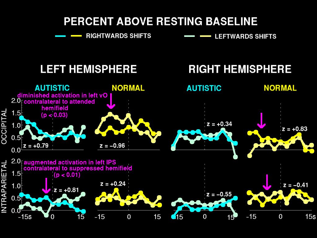
In our fMRI analysis of autism, we again find that ventral occipital cortex in both hemispheres is somewhat more active in response to targets in the left hemifield than to targets in the right. Thus, here again, the hemispheres fail to activate independently of each other.
This result replicates our earlier EEG finding, using a different technique and a separate group of subjects.
Complementing this reduced attention effect in ventral occipital cortex is an augmented attention effect in intraparietal sulcus contralateral to the suppressed hemifield.
For the group comparison, this effect reached significance in left intraparietal cortex.
It's also worth noting that the effect in right intraparietal cortex was significant within the two male subjects whose clinical diagnosis was autism.
In the controls, in contrast, the intraparietal measure was not significant within any of the individual subjects; it emerged only as a group effect.